
Lesions Exhibiting Necrobiosis
There are three dermatological conditions which, when viewed under the microscope, are characterized by the presence of degenerative collagen (necrobiosis). These include granuloma annulare, rheumatoid nodules, and necrobiosis lipoidica diabeticorum. These lesions can usually be differentiated on a clinical basis. Normally, granuloma annulare appears as a predominately dermal (skin) lesion. The rheumatoid nodule is a nodule (lump) arising in the subcutaneous tissue over the surfaces of the fingers, elbows, knees, and toes. Necrobiosis lipoidica diabeticorum will show extensive loss of tissue substance of the skin on the shins of a patient with diabetes. While the clinical characteristics of these three lesions are quite distinctive, in some cases there is a significant overlap.
Let’s look at each of these lesions individually
Granuloma Annulare
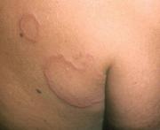
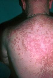
These lesions are common in children as well as young adults. They are particularly common in those individuals with a strong family history of diabetes mellitis. The initial granuloma annulare lesion often resembles a mosquito bite and appears on the wrist, ankle, elbow, knee, or the digit of a hand or foot. These lesions are asymptomatic and do not itch. A granuloma annulare lesion gradually spreads outward in a circular pattern with central clearing and no apparent scaling.
In most cases there are only one or two lesions, but dozens of lesions could develop. In most cases, GA can be easily cleared with topical or intralesional steroid therapy. However, there are occasions when this condition can be widespread and there may forms of systemic therapy including UVB therapy, dapsone, methyltrexate and retinoid therapy that can be effective in clearing these more severe lesions. See the examples of Granuloma Annulare shown below.
Rheumatoid Nodules
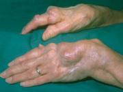
As you would expect by the name, these lesions normally occur in patients with rheumatoid arthritis. The lesions are usually asymptomatic and are most often located over the extensor surfaces of joints on the hands, wrists, elbows, knees, ankles, and toes.
Necrobiosis Lipoidica Diabeticorum
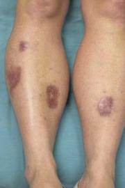
Clinically, this very distinctive lesion can occur on many parts of the body, but normally it is seen on the pretibial (shin) area of the lower legs. It is generally tied to diabetes, and may appear before the onset of the disease, in its early stages, or in the chronic stages of the disease. These lesions have three distinct clinical characteristics. The first is significant atrophy or loss of tissue substance. The growth of the lesion often results in the destruction of hair follicles and sebaceous glands. The second characteristic is a thinning of the epidermis. Telangiectatic vessels (small bright red blood vessels) may become visible, particularly around the periphery of the lesions. The third sign is a yellowish or orange color in the central portion of the lesion caused by lipid deposits. Necrobiosis lipoidica diabeticorum lesions may become quite extensive and involve almost the entire cutaneous surface of the pretibial area. If left untreated, they can become extensive ulcerated lesions which are extremely resistant to conventional therapy. Intralesional steroid therapy in the early stages of this problem can be quite effective. However, as with any lesion associated with diabetes, careful management of the blood glucose level is essential to create an environment where this lesion can be effectively treated.